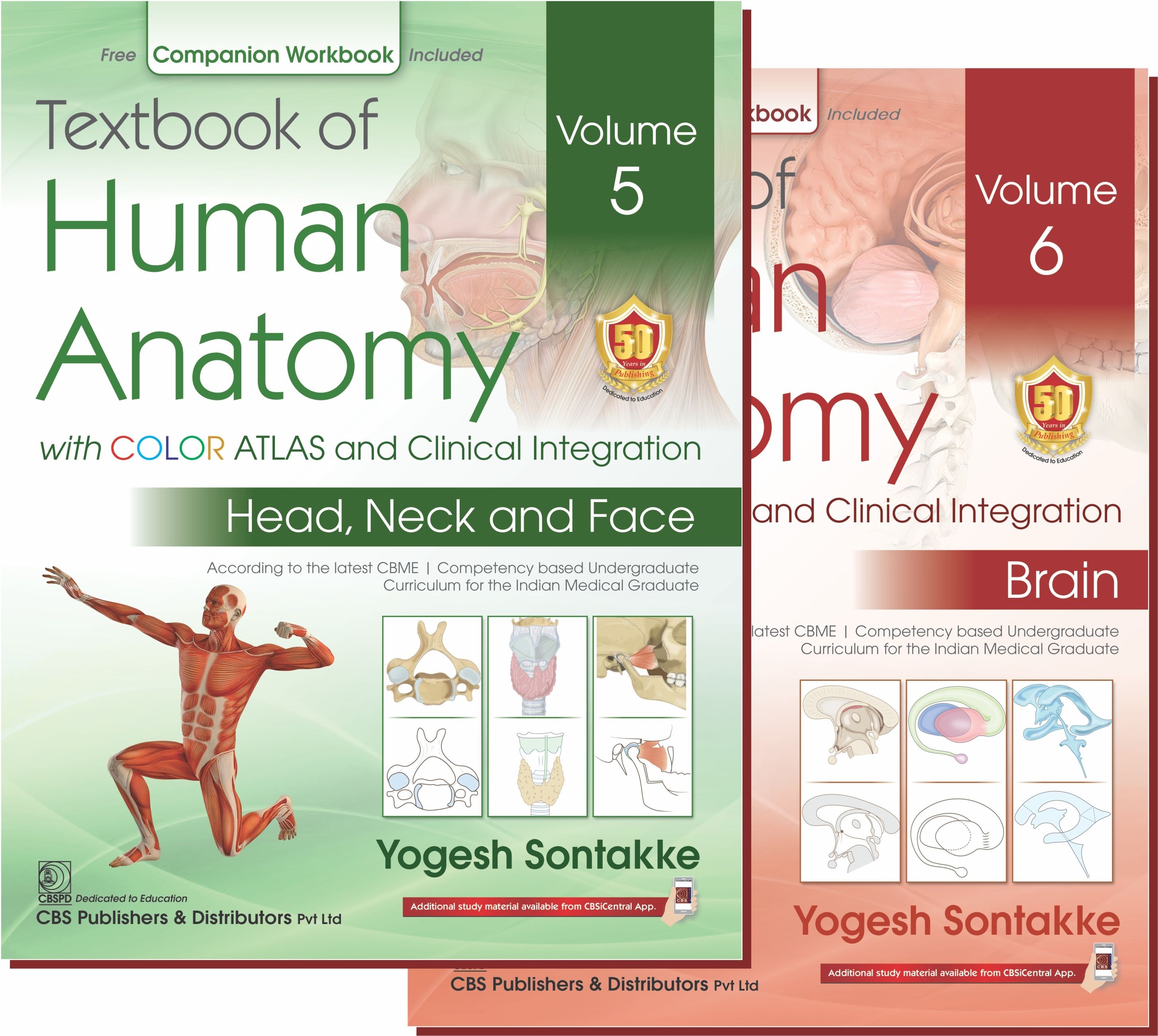
Textbook of Human Anatomy with COLOR ATLAS and Clinical Integration Volume 5 Head ,Neck and Face ;Volume 6 Brain(2 Volume Set)
in Medical
Created by
Yogesh Sontakke
About this course
Digital Content of Textbook of Human Anatomy is written by Dr. Yogesh Sontakke and Dr Ujwala Bhanarkar – authors of many Best-seller books. This book helps students to fulfil the
requirements as per the latest CBME Guidelines and gives the clinical orientation. The book has concise text with functional correlation, 2350 plus Beautiful Color atlas illustrations,
1050 Line diagrams, 450 Clinical images, 620 Flowcharts, 210 Boxes on important topics and markings for NeXT, MCQ, Viva voce, and Clinical facts;.
Comments (0)
PDF
63 Parts
Exterior of Skull
0.92 MB
Interior of Skull
0.22 MB
Cervical Vertebrae
1.11 MB
Mandible
1.04 MB
Scalp and Temple
0.73 MB
Face
0.74 MB
Eyelids and Lacrimal Apparatus
1.06 MB
Neck: Skin, Fascia and Sternocleidomastoid
0.71 MB
Neck: Posterior Triangle
1.13 MB
Neck: Anterior Triangle
0.54 MB
Parotid Region
0.53 MB
Temporal and Infratemporal Regions
0.71 MB
Temporomandibular Joint and Styloid Apparatus
0.95 MB
Submandibular Region
0.68 MB
Thyroid and Parathyroid Glands
0.55 MB
Trachea, Esophagus and Thymus
0.73 MB
Neck: Blood Vessels and Lymphatics
0.55 MB
Last Four Cranial Nerves
0.78 MB
Peripheral Parasympathetic Ganglia and Cervical Sympathetic Chain
0.67 MB
Prevertebral and Paravertebral Regions
1.85 MB
Back of the Neck
0.87 MB
Joints of the Neck
1.09 MB
Cranial Cavity
0.47 MB
Pituitary Gland
0.5 MB
Orbit I: Bony Orbit and Muscles of Orbit
1.78 MB
Orbit II: Nerves and Vessels of Orbit
0.68 MB
Mouth (Oral Cavity)
1.97 MB
Tongue and Taste
0.47 MB
Palate
0.67 MB
Pharynx
0.61 MB
Palatini Tonsils and Auditory Tube
1.1 MB
Nose
1.42 MB
Paranasal Air Sinuses and Pterygopalatine Fossa
1.15 MB
Larynx
1.99 MB
External and Middle Ear
1.87 MB
Internal Ear
0.51 MB
Eyeball
0.46 MB
Surface Marking of Head, Neck and Face
1.93 MB
Radiological Anatomy of Head, Neck and Face
1.58 MB
Introduction
1.48 MB
Peripheral Nerves and Ganglia
0.42 MB
Receptors
0.38 MB
Spinal Cord
0.69 MB
Tracts of Spinal Cord
1.62 MB
Medulla Oblongata
0.49 MB
Pons
1.38 MB
Midbrain
1.78 MB
Cerebellum
1.6 MB
Fourth Ventricle
0.89 MB
Diencephalon I: Thalamus, Epithalamus, Metathalamus
1.79 MB
Diencephalon II: Hypothalamus
1.19 MB
Third Ventricle
0.71 MB
Cerebrum
0.62 MB
Basal Ganglia
1.2 MB
White Matter of Cerebrum
0.57 MB
Limbic System
1.71 MB
Lateral Ventricle
0.85 MB
Reticular Formation
0.96 MB
Blood Supply of Brain
0.6 MB
Meninges and Cerebrospinal Fluid
1.33 MB
Automatic Nervous System
1.05 MB
Cranial Nerves
0.54 MB
Surface Anatomy and Imaging of Central Nervous System
1.93 MB
Videos
201 Parts
Figure 1.4. Norma Verticalis
-
Figure 1.6. Norma Occipitalis
-
Figure 1.9. Norma Frontalis
-
Figure 1.10. Norma Frontalis: Attachments
-
Figure 1.13. Norma Lateralis
-
Figure 1.14. Norma Lateralis: Attachments
-
Figure 1.20. Norma Basalis
-
Figure 1.22. Norma Basalis: Attachments
-
Figure 1.26. Foramen Lacerum
-
Figure 1.27. Foramen Magnum
-
Figure 1.28. Jugular Foramen
-
Figure 1.29. Fetal Skull
-
Figure 2.7. Features of Cranial Fossae
-
Figure 2.8. Superior Orbital Fissure and Optic Canal
-
Figure 3.3. Typical Cervical Vertebra
-
Figure 3.6. Atlas Vertebra
-
Figure 3.7. Relations of Atlas Vertebra
-
Figure 3.9. Axis Vertebra
-
Figure 3.11. Seventh Cervical Vertebra
-
Figure 3.13. Hyoid Bone
-
Figure 3.14. Hyoid Bone: Attachments
-
Figure 4.5. Mandible
-
Figure 4.6. Mandible: Attachments
-
Figure 4.7. Relations of the mandible
-
Figure 5.3. Layers of Scalp
-
Figure 5.6. Scalp: Arterial Supply
-
Figure 5.7. Scalp: Nerve Supply
-
Figure 6.6. Muscles of Facial Expression
-
Figure 6.10. Muscles of Facial Expression: Motor Supply
-
Figure 6.11. Sensory Innervation of the Face
-
Figure 6.12. Face: Sensory Nerves
-
Figure 6.14. Face: Arterial Supply
-
Figure 6.16. Face: Venous Drainage
-
Figure 6.18. Scalp and Face: Lymphatic Drainage
-
Figure 7.2. Structure of the Eyelid
-
Figure 7.12. Lacrimal Apparatus
-
Figure 7.14. Innervation of Lacrimal Gland
-
Figure 8.4. Fibers of Platysma
-
Figure 8.5. Cutaneous Nerves of Neck
-
Figure 8.6. Superficial Veins of Neck
-
Figure 8.7. Superficial Cervical Lymph Nodes
-
Figure 8.8. Superficial Cervical Lymph Nodes
-
Figure 8.14. Supraclavicular Space
-
Figure 8.17. Right Carotid Sheath
-
Figure 8.18. Retropharyngeal Space
-
Figure 9.5. Posterior Triangle of Neck: Boundaries
-
Figure 9.7. Roof of Posterior Triangle
-
Figure 9.9. Posterior Triangle: Boundaries and Floor
-
Figure 9.10. Posterior Triangle of Neck: Divisions
-
Figure 9.12. Posterior Triangle of Neck: Contents
-
Figure 10.5. Anterior Triangle: Boundaries and Subdivisions
-
Figure 10.7. Submental Triangle: Boundaries and Contents
-
Figure 10.9. Digastric Triangle: Boundaries
-
Figure 10.10. Digastric Triangle: Contents
-
Figure 10.13. Carotid Triangle: Boundaries and Floor
-
Figure 10.14. Contents of Carotid Triangle and Posterior Part of Digastric Triangle
-
Figure 10.16. Infrahyoid Muscles
-
Figure 10.17. Ansa Cervicalis: Formation and Distribution
-
Figure 10.18. External Carotid Artery: Branches
-
Figure 11.1. Parotid Bed and Location of Parotid Gland
-
Figure 11.2. Parotid Capsule
-
Figure 11.3. External Features of Parotid Gland
-
Figure 11.4. Relations of Parotid Gland
-
Figure 11.6. Structures Emerging at Periphery of Parotid Gland
-
Figure 11.7. Structures Traversing Parotid Gland
-
Figure 11.10. Innervation of Parotid Glane
-
Figure 12.5. Masseter Muscle: Origin and Insertion
-
Figure 12.6. Temporalis Muscle: Origin and Insertion
-
Figure 12.8. Medial Pterygoid Muscle: Origin and Insertion
-
Figure 12.10. Lateral Pterygoid Muscle: Origin and Insertion
-
Figure 12.11. Lateral Pterygoid Muscle: Relations
-
Figure 12.12. Medial Pterygoid Muscle: Superficial Relations
-
Figure 12.13. Maxillary Artery: Branches
-
Figure 12.15. Mandibular Nerve: Course and Distribution
-
Figure 12.16. Inferior Alveolar Nerve
-
Figure 12.17. Inferior Alveolar Nerve Block
-
Figure 12.18. Course of Chorda Tympani Nerve
-
Figure 12.19. Chorda Tympani Nerve: Functional Components
-
Figure 12.20. Otic Ganglion
-
Figure 13.3. Temporomandibular Joint: Articular Surfaces
-
Figure 13.4. Articular Disc of Temporomandibular Joint
-
Figure 13.6. Ligaments of Temporomandibular Joint
-
Figure 13.7. Sphenomandibular Ligament: Superficial Relations
-
Figure 13.8. Relations of the Sphenomandibular Ligament
-
Figure 13.13. Styloid Apparatus
-
Figure 14.2. Digastric Muscle
-
Figure 14.3. Relations of Posterior Belly of Digastric Muscle
-
Figure 14.4. Stylohyoid muscle
-
Figure 14.6. Mylohyoid Muscle
-
Figure 14.7. Geniohyoid and Hyoglossus Muscles
-
Figure 14.8. Superficial Relations of Hyoglossus Muscle
-
Figure 14.9. Styloglossus Muscle
-
Figure 14.10. Genioglossus Muscle
-
Figure 14.11. Parts of Submandibular Gland
-
Figure 14.12. Capsule of Superficial Part of Submandibular Gland
-
Figure 14.13. Relations of Submandibular Gland
-
Figure 14.14. Relations of Superficial part of submandibular gland
-
Figure 14.15. Relations of Medial Surface of Submandibular Gland
-
Figure 14.16. Deep Part of Submandibular Gland and Sublingual Gland
-
Figure 14.17. Submandibular Ganglion and its Connections
-
Figure 15.3. Thyroid Gland: Parts and Extent
-
Figure 15.4. Capsules of Thyroid and Prostate Glands
-
Figure 15.5. Parts of Lobes of Thyroid Gland
-
Figure 15.6. Relations of Thyroid Gland
-
Figure 15.7. Thyroid Gland: Relations of Medial Surface of Lateral Lobe
-
Figure 15.8. Arterial Supply of Thyroid Gland
-
Figure 15.9. Venous Drainage of Thyroid Gland
-
Figure 15.10. Thyroid Gland: Lymphatic Drainage
-
Figure 17.1. Beginning of Subclavian Arteries
-
Figure 17.5. Branches of Subclavian Arteries
-
Figure 17.6. Course and parts of Vertebral Artery
-
Figure 17.10. Costocervical and Thyrocervical Trunk
-
Figure 17.12. Internal Carotid Artery
-
Figure 17.13. Subclavian and Brachiocephalic Veins
-
Figure 17.14. Internal Jugular Veins
-
Figure 17.16. Cervical Lymph Nodes
-
Figure 18.5. Glossopharyngeal Nerve: Course and Distribution
-
Figure 18.6. Gag Reflex
-
Figure 18.7. Vagus Nerve: Functional Components and Nuclei
-
Figure 18.8. Extracranial Course of Last Three Cranial Nerves
-
Figure 18.10. Distribution of Vagus Nerve in Head and Neck
-
Figure 18.13. Functional Components and Nuclei of Accessory Nerve
-
Figure 18.15. Functional Components and Nucleus of Hypoglossal Nerve
-
Figure 18.16. Hypoglossal Nerve
-
Figure 18.17. Course and Distribution of Hypoglossal Nerve
-
Figure 19.3. Right Cervical Sympathetic Trunk and its Branches
-
Figure 20.13. Cervical Plexus
-
Figure 20.15. Phrenic Nerve
-
Figure 21.17. Suboccipital Muscles
-
Figure 21.18. Boundaries and Contents of Suboccipital Triangle
-
Figure 21.20. Suboccipital Nerve
-
Figure 22.5. Median Atlantoaxial Articulation
-
Figure 22.6. Ligaments of Cervical Vertebrae
-
Figure 23.8. Cavernous Sinuses
-
Figure 23.10. Tributaries and Communications of Cavernous Sinus
-
Figure 23.11. Communications of Cavernous Sinus
-
Figure 24.2. Parts of Hypophysis Cerebri
-
Figure 25.1. Superior Orbital Fissure and Optic Canal
-
Figure 25.13. Nerve Supply of Extraocular Muscles
-
Figure 25.14. Actions of Extraocular Muscles
-
Figure 26.1. Functional Components and Nuclei of Oculomotor Nerve
-
Figure 26.5. Oculomotor Nerve
-
Figure 26.6. Oculomotor Nerve
-
Figure 26.8. Trochlear Nucleus and Beginning of Trochlear Nerve
-
Figure 26.10. Trochlear Nerve
-
Figure 26.11. Trochlear and Abducent Nerves
-
Figure 26.12. Abducent Nucleus and Beginning of Abducent Nerve
-
Figure 26.14. Branches of Ophthalmic Nerve
-
Figure 26.15. Ciliary Ganglion
-
Figure 26.16. Ophthalmic Artery
-
Figure 27.11. Nerve Supply of Teeth
-
Figure 28.2. Dorsal Surface of Tongue
-
Figure 28.8. Extrinsic Muscles of Tongue
-
Figure 28.9. Muscles of Tongue
-
Figure 28.12. Arterial Supply of Tongue
-
Figure 28.13. Lymphatic Drainage of Tongue
-
Figure 28.14. Nerve Supply of Tongue with Embryological Source
-
Figure 29.2. Oral Aspect of Hard Palate
-
Figure 29.4. Muscles of Soft Palate
-
Figure 29.7. Passavants Ridge
-
Figure 30.1A. Subdivisions of Pharynx
-
Figure 30.3. Pharynx
-
Figure 30.4. Nasopharynx
-
Figure 30.6. Boundaries of the Oropharyngeal Isthmus
-
Figure 30.7. Piriform Fossa
-
Figure 30.8. Structure of Pharyngeal wall
-
Figure 30.10. Constrictor Muscles of Pharynx
-
Figure 30.11. Structures Passing Through Gap in Wall of Pharynx
-
Figure 30.12. Structures Passing Through Gaps in Pharyngeal Wall
-
Figure 30.14. Longitudinal Muscles of Pharynx
-
Figure 31.1. Waldeyers Ring
-
Figure 31.5. Relations of Palatine Tonsil
-
Figure 31.7. Arteries Supplying Palatine Tonsil
-
Figure 31.14. Pharyngotympanic Tube
-
Figure 32.5. Formation of Nasal Septum
-
Figure 32.6. Arterial Supply of Nasal Septum
-
Figure 32.7. Nerve Supply of Nasal Septum
-
Figure 32.10. Features of Lateral Wall of Nasal Cavity
-
Figure 32.12. Lateral Wall of the Nose Showing Openings
-
Figure 32.13. Arterial Supply of Lateral Wall of Nose
-
Figure 32.14. Nerve Supply of Lateral Wall of Nose
-
Figure 33.8. Boundaries and Communications of Pterygopalatine Fossa
-
Figure 33.9. Maxillary Nerve
-
Figure 33.10. Pterygopalatine ganglion
-
Figure 33.11. Branches of Pterygopalatine Ganglion
-
Figure 34.5. Cartilages and Skeleton of Larynx
-
Figure 34.15. Ligaments and Membranes of Larynx
-
Figure 34.17. Intrinsic Muscles of Larynx
-
Figure 34.18. Actions of Intrinsic Muscles of Larynx
-
Figure 34.23. Cavity of Larynx
-
Figure 35.8. Layers of Tympanic Membrane
-
Figure 35.9. External Surface of Tympanic Membrane
-
Figure 35.13. Internal Surface of Tympanic Membrane
-
Figure 35.16. Walls of Middle Ear
-
Figure 35.17. Walls of Middle Ear
-
Figure 35.18. Medial Wall of Middle Ear
-
Figure 35.20. Ear Ossicles
-
Figure 36.2. Right Bony Labyrinth
-
Figure 37.2. Structure of Eyeball
-
Figure 37.5. Layers of Retina and Ultrastructure of Rod and Cone
-
Figure 37.9. Basic Components of Visual Pathway
-

0
0 Reviews