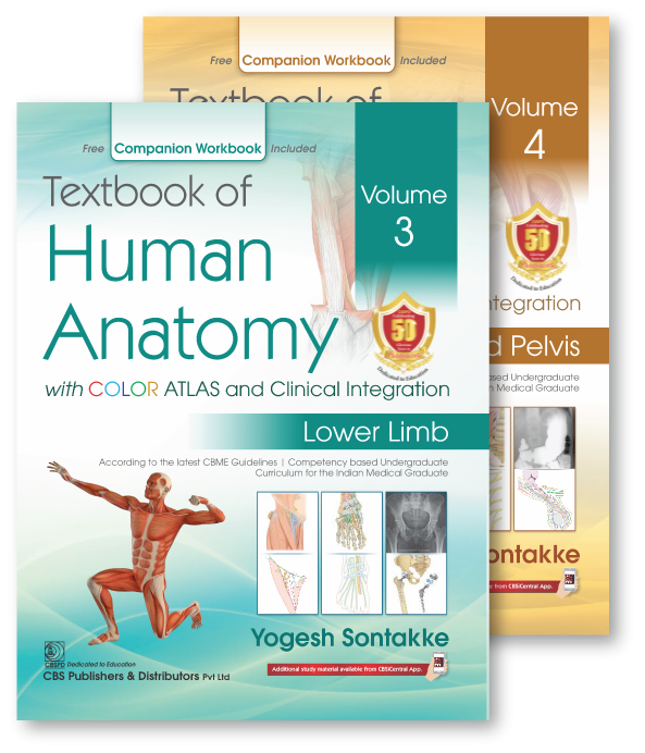
Textbook of Human Anatomy with COLOR ATLAS and Clinical Integration Volume 3 (Lower Limb) & 4 (Abdomen and Pelvis)
in Medical
Created by
Yogesh Sontakke
About this course
Digital Content of Textbook of Human Anatomy is written by Dr. Yogesh Sontakke and Dr Ujwala Bhanarkar – authors of many Best-seller books. This book helps students to fulfil the
requirements as per the latest CBME Guidelines and gives the clinical orientation. The book has concise text with functional correlation, 2350 plus Beautiful Color atlas illustrations,
1050 Line diagrams, 450 Clinical images, 620 Flowcharts, 210 Boxes on important topics and markings for NeXT, MCQ, Viva voce, and Clinical facts;.
Comments (0)
PDF
27 Parts
Chapter- 1 Introduction to Lower limb
0.05 MB
Chapter- 2 Hip Bone
0.7 MB
Chapter- 3 Bone of Thigh: Femur
0.54 MB
Chapter- 4 Bones of Leg: Tibia and Fibula
0.71 MB
Chapter- 5 Front of the Thigh I: Fascia and Muscles
0.56 MB
Chapter- 6 Front of the Thigh I: Fascia and Muscles
0.57 MB
Chapter- 7 Front of the Thigh II: Femoral Triangle, Nerves and Vessels
0.93 MB
Chapter- 8 Medial Side of Thigh
0.24 MB
Chapter- 9 Adductor Canal
0.24 MB
Chapter- 10 Gluteal Region
0.91 MB
Chapter- 11 Posterior Compartment of Thigh
0.65 MB
Chapter- 12 Hip Joint
0.33 MB
Chapter- 13 Popliteal Fossa
0.67 MB
Chapter- 14 Front of Leg and Dorsum of Foot
1.21 MB
Chapter- 15 Lateral and Medial Sides of Leg
0.35 MB
Chapter- 16 Back of the Leg
0.71 MB
Chapter- 17 Knee Joint
0.36 MB
Chapter- 18 Sole of the Foot
0.88 MB
Chapter- 19 Ankle and Tibiofibular Joints
0.41 MB
Chapter- 20 Joints of Foot
0.18 MB
Chapter- 21 Arches of Foot
0.62 MB
Chapter- 22 Cutaneous Innervation, Venous Drainage, and Lymphatic Drainage of Lower Limb
0.85 MB
Chapter- 23 Major Nerves of Lower Limb
1.08 MB
Chapter- 24 Surface Anatomy of Lower Limb
1.05 MB
Chapter- 25 Radiological Anatomy of Lower Limb
0.85 MB
Chapter- 26 Development of Lower Limb
0.24 MB
Chapter- 27 Functional Anatomy of Lower Limb: Weight Transmission, Walking Cycle, and Functional Correlation of Limbs
0.28 MB
Videos
142 Parts
Lower Limb Figure 2.3. Schematic Representation of Hip Bone
-
Lower Limb Figure 2.5. Attachments of Hip Bone
-
Lower Limb Figure 2.6. Attachments of Hip Bone
-
Lower Limb Figure 2.7. Attachments of Iliac Crest
-
Lower Limb Figure 3.3. Femur
-
Lower Limb Figure 3.6. Attachments of Femur
-
Lower Limb Figure 3.8. Neck-Shaft Angle of Femur
-
Lower Limb Figure 3.9. Angle of Femoral Torsion
-
Lower Limb Figure 3.10. Trochanter-Shaft Angle of Femur
-
Lower Limb Figure 3.11. Blood Supply of Head of Femur
-
Lower Limb Figure 3.13. Attachments of Linea Aspera
-
Lower Limb Figure 3.16. Fractures of Femur
-
Lower Limb Figure 3.17. Coxa Vara and Coxa Valga
-
Lower Limb Figure 3.19. Features of Patella
-
Lower Limb Figure 3.20. Attachments of Patella
-
Lower Limb Figure 4.2. Tibia
-
Lower Limb Figure 4.4. Superior view of Tibia
-
Lower Limb Figure 4.5. Attachments of Tibia
-
Lower Limb Figure 4.13. Fibula
-
Lower Limb Figure 4.16. Attachments of the Surfaces of Fibula
-
Lower Limb Figure 4.17. Lower end of Fibula
-
Lower Limb Figure 5.2. Bones of the Foot
-
Lower Limb Figure 5.4. Talus
-
Lower Limb Figure 5.5. Attachments of Talus
-
Lower Limb Figure 5.6. Attachments of Bones of Foot
-
Lower Limb Figure 5.9. Calcaneus
-
Lower Limb Figure 5.10. Attachments of Calcaneus
-
Lower Limb Figure 6.2. Midinguinal Point and Midpoint of Inguinal Ligament
-
Lower Limb Figure 6.3. Holden's Line
-
Lower Limb Figure 6.4. Cutaneous Nerves on the Front of the Thigh
-
Lower Limb Figure 6.6. Superficial Vessels of Thigh
-
Lower Limb Figure 6.7. Superficial Inguinal Lymph Nodes
-
Lower Limb Figure 6.8. Iliotibial Tract
-
Lower Limb Figure 6.11. Compartments of Thigh
-
Lower Limb Figure 6.13. Muscles of Anterior Compartment of Thigh
-
Lower Limb Figure 7.2. Boundaries of Femoral Triangle
-
Lower Limb Figure 7.4. Muscles Forming the Floor of Femoral Triangle
-
Lower Limb Figure 7.6. Contents of Femoral Triangle
-
Lower Limb Figure 7.7. Contenet of Femoral Triangle
-
Lower Limb Figure 7.9. Formation of the Femoral Sheath
-
Lower Limb Figure 7.10. Femoral Sheath
-
Lower Limb Figure 7.11. Transverse Section of the Femoral Sheath with its Contents
-
Lower Limb Figure 7.12. Femoral Hernia
-
Lower Limb Figure 7.13. Abberent Obturator Artery
-
Lower Limb Figure 7.15. Relations of the Femoral Artery
-
Lower Limb Figure 7.16. Branches of Femoral Artery
-
Lower Limb Figure 7.23. Femoral Nerve
-
Lower Limb Figure 8.6. Obturator Nerve
-
Lower Limb Figure 8.7. Branches of Obturator Nerve
-
Lower Limb Figure 8.8. The Course and Distribution of the Accessory Obturator Nerve
-
Lower Limb Figure 9.2. Walls of Adductor Canal
-
Lower Limb Figure 9.3. Contents of Adductor Canal
-
Lower Limb Figure 9.4. Contents of Adductor Canal
-
Lower Limb Figure 9.5. Relationship of Femoral Artery and Vein
-
Lower Limb Figure 10.9. Gluteus maximus muscle
-
Lower Limb Figure 10.13. Muscles of Gluteal region
-
Lower Limb Figure 10.16. Trochanteric Anastomosis
-
Lower Limb Figure 10.17. Cruciate Anastomosis
-
Lower Limb Figure 10.21. Structures in the Gluteal region under cover of gluteus maximus
-
Lower Limb Figure 11.5. Semitendinosus, semimembranosus, and biceps femoris muscles
-
Lower Limb Figure 11.9. Formation of sciatic nerve
-
Lower Limb Figure 11.11. Relations of sciatic nerve
-
Lower Limb Figure 11.12. Branches of sciatic nerve
-
Lower Limb Figure 11.15. Anastomosis on the back of the thigh
-
Lower Limb Figure 12.2. Hip joint: articular surfaces
-
Lower Limb Figure 12.3. Attachment of capsule of hip joint on femur
-
Lower Limb Figure 12.5. Iliofemoral and pubofemoral ligaments
-
Lower Limb Figure 12.7. Iliofemoral and ischiofemoral ligaments
-
Lower Limb Figure 12.9. Relations of hip joint
-
Lower Limb Figure 12.12. Blood Supply of Head of Femur
-
Lower Limb Figure 13.2. Boundaries of popliteal fossa
-
Lower Limb Figure 13.3. Structures in the roof of popliteal fossa
-
Lower Limb Figure 13.4. Floor of popliteal fossa
-
Lower Limb Figure 13.6. Floor of popliteal fossa
-
Lower Limb Figure 13.7. Boundaries of popliteal fossa
-
Lower Limb Figure 13.8. Branches of popliteal artery
-
Lower Limb Figure 13.13. Branches of tibial nerve in popliteal fossa and leg
-
Lower Limb Figure 13.14. Branches of common peroneal nerve in popliteal fossa and leg
-
Lower Limb Figure 13.15. Arterial anastomosis around the knee
-
Lower Limb Figure 14.1. Osseo-fascial compartments of leg
-
Lower Limb Figure 14.2. Surface landmarks on the leg and dorsum of foot
-
Lower Limb Figure 14.3. Superficial veins of the front of the leg and dorsum of the foot
-
Lower Limb Figure 14.4. Cutaneous nerves on the front of the leg and dorsum of the foot
-
Lower Limb Figure 14.6. Superior and inferior extensor retinaculum on the front of the ankle
-
Lower Limb Figure 14.12. Extensor digitorum longus
-
Lower Limb Figure 14.16. Relations of anterior tibial artery and deep peroneal nerve
-
Lower Limb Figure 14.17. Anterior tibial artery
-
Lower Limb Figure 14.18. Course and distribution of deep peroneal nerve on the dorsum of the foot
-
Lower Limb Figure 14.19. Injury to the deep peroneal nerve
-
Lower Limb Figure 14.20. Sensory innervation of the dorsum of the foot
-
Lower Limb Figure 14.22. Extensor digitorum brevis and extensor hallucis brevis
-
Lower Limb Figure 14.23. Course and distribution of dorsalis pedis artery
-
Lower Limb Figure 15.1. Lateral compartment of leg and its content
-
Lower Limb Figure 15.6. Peroneus brevis
-
Lower Limb Figure 16.1. Contents of posterior compartment of leg, section passing through the midle of the leg
-
Lower Limb Figure 16.2. Surface landmarks of back of leg
-
Lower Limb Figure 16.3. Cutaneous innervation of back of leg
-
Lower Limb Figure 16.4. Cutaneous innervation of back of leg
-
Lower Limb Figure 16.6. Flexor retinaculum
-
Lower Limb Figure 16.8. Gastrocnemius, soleus, and plantaris
-
Lower Limb Figure 16.14. Tibialis posterior, flexor digitorum longus, flexor hallucis longus, popliteus
-
Lower Limb Figure 16.18. Branches of posterior tibial artery
-
Lower Limb Figure 16.21. Branches of tibial nerve in popliteal fossa and leg
-
Lower Limb Figure 17.6. Superior view of tibia
-
Lower Limb Figure 17.9. Divisions of joint cavity
-
Lower Limb Figure 17.14. Relations of knee joint
-
Lower Limb Figure 17.15. Mechanism of locking and unlocking of knee
-
Lower Limb Figure 18.1. Transverse section of right foot through metatarsals showing the arrangement of structures in the sole
-
Lower Limb Figure 18.2. Cutaneous innervation of the sole of foot and dermatomes
-
Lower Limb Figure 18.5. Plantar aponeurosis
-
Lower Limb Figure 18.7. Muscles of first layer of sole
-
Lower Limb Figure 18.8. Muscles of second layer of sole
-
Lower Limb Figure 18.10. Muscles of third layer of sole
-
Lower Limb Figure 18.12. Palmar interossei of foot
-
Lower Limb Figure 18.14. Dorsal interossei of foot
-
Lower Limb Figure 18.16. Medial plantar nerve
-
Lower Limb Figure 18.17. Lateral plantar nerve
-
Lower Limb Figure 18.19. Medial and Lateral Plantar arteries
-
Lower Limb Figure 18.20. Proximal and distal perforating arteries
-
Lower Limb Figure 19.3. Ankle joint: articular surfaces
-
Lower Limb Figure 19.4. Ligaments and articular surfaces of ankle joint
-
Lower Limb Figure 19.5. Practice figure: Deltoid ligament
-
Lower Limb Figure 19.6. Medial plantar nerve
-
Lower Limb Figure 19.7. Lateral plantar nerve
-
Lower Limb Figure 20.4. Midtarsal joint
-
Lower Limb Figure 20.10. Axis of inversion and eversion
-
Lower Limb Figure 21.1. Anatomical and Functional Segments of Skeleton of Foot
-
Lower Limb Figure 21.5. Bones forming the longitudinal arches of foot
-
Lower Limb Figure 21.6. Longitudinal arches of foot
-
Lower Limb Figure 21.7. Ligaments maintaining medial longitudinal arch
-
Lower Limb Figure 21.8. Factors maintaining the longitudinal arches
-
Lower Limb Figure 21.9. Ligaments maintaining lateral longitudinal arch
-
Lower Limb Figure 21.10. Tendons of peroneus longus, peroneus brevis, and tibialis anterior
-
Lower Limb Figure 22.1. Cutaneous innervation of front of lower limb
-
Lower Limb Figure 22.2. Cutaneous innervation of gluteal region
-
Lower Limb Figure 22.3. Cutaneous innvervation of back of leg
-
Lower Limb Figure 22.4. Cutaneous innvervation of sole of the foot
-
Lower Limb Figure 22.8. Veins of lower limb
-
Lower Limb Figure 22.9. Superficial veins of the front of the leg and dorsum of the foot
-
Lower Limb Figure 22.10. Long Saphenous vein and its tributaries
-
Lower Limb Figure 22.14. Superficial Inguinal Lymph Nodes
-
Lower Limb Figure 15.4. Peroneus longus
-

0
0 Reviews